Bones In Leg Diagram / Pin By Genna Hornsby On Anatomy Human Anatomy And Physiology Medical Anatomy Leg Anatomy. These muscles work together to produce movements such as standing, walking, running, and jumping. The calcaneus, or heel bone : To explain the term in layman's language, it is the heel bone in the skeletal system. The lacrimal bone is the smallest bone of the face, and there are 2 such bones, each one forming a part of the median wall of the orbital cavity. Tendons connect the knee bones to the leg muscles that move the knee.
The bones of the leg and foot form part of the appendicular skeleton that supports the many muscles of the lower limbs. The tarsal bones in the foot are located amongst tibia, metatarsal bones, and fibula. The bones shown in the chest and hip region in the labeled human skeleton diagram are the ribs, vertebrae, pelvis, os coxae, sacrum and coccyx. To explain the term in layman's language, it is the heel bone in the skeletal system. These are 2 oblong bones, and their size varies in different individuals.
Foot Bones Anatomy Conditions And More from cdn-prod.medicalnewstoday.com The patella is the kneecap and articulates with the distal femur. Also called the shin bone, the tibia is the longer of the two bones in the. These are the femur, patella, tibia, fibula, tarsal bones, metatarsal bones, and phalanges (see figure 6.51). The pubis, ischium, and ilium together constitute the pelvis while the thigh bone is the femur. Many muscles that move the trunk and legs, such as our abdominal muscles, attach to the hip bones. The bones shown in the chest and hip region in the labeled human skeleton diagram are the ribs, vertebrae, pelvis, os coxae, sacrum and coccyx. The talus, or ankle bone: At the same time, the bones and joints of the leg and foot must be strong enough to support the body's weight while remaining.
The calcaneus is largest of the.
The lower limb contains 30 bones. Related posts of bones leg diagram picture bone anatomy of upper extremity. To explain the term in layman's language, it is the heel bone in the skeletal system. The smaller bone that runs alongside the tibia (fibula) and the kneecap (patella) are the other bones that make the knee joint. Also called the shin bone, the tibia is the longer of the two bones in the. The femur is the single bone of the thigh. It connects with the tibia and fibula bones of the lower leg. The bones of the hip include the femur, the ilium, the ischium, and the pubis. Bone anatomy of upper extremity 12 photos of the bone anatomy of upper extremity anatomy of upper limb bones pdf, bone anatomy of the upper limb, bone anatomy of upper extremity, bone anatomy of upper limb ppt, skeletal anatomy of the upper extremity, bone, anatomy. Depending on the cause, leg lumps may be single or while these conditions are rare, both benign and malignant tumors of the skin, soft tissues, or bones can sometimes feel. The nasal bones are two in number, and together form the bridge of the nose. Another bone that is part of the lower leg and the knee joint is called the fibula.this is a bone located on the lateral, or outer part, of the lower leg and is more commonly known as the calf bone. This image is an edited version of this image that was created by user:ladyofhats (mariana ruiz villarreal).
The femur is the single bone of the thigh. A leg bone is a bone found in the leg.these can include any the following: The pubis, ischium, and ilium together constitute the pelvis while the thigh bone is the femur. The tibia and fibula are two long bones that run parallel to each other, forming the scaffold of the leg and providing attachment points for many muscles. Another bone that is part of the lower leg and the knee joint is called the fibula.this is a bone located on the lateral, or outer part, of the lower leg and is more commonly known as the calf bone.
Leg Fracture What You Need To Know from www.drugs.com The bones of the leg and foot form part of the appendicular skeleton that supports the many muscles of the lower limbs. The tibia and the fibula, at the top of the ankle joint. The calcaneus, or heel bone : In addition, the broad hip bones provide protection to the delicate internal organs of the pelvis, such as the intestines, urinary bladder, and uterus. Bone diagram forehead (frontal bone) nose bones (nasals) cheek bone (zygoma) upper jaw (maxilla) lower jaw (mandible) breast bone (sternum) upper arm bone (humerus) lower arm bone (ulna) thigh bone (femur) collar bone (clavicle) toe bones (phalanges) ankle bones (tarsals) kneecap (patella) shin bone The thigh bone, or femur, is the large upper leg bone that connects the lower leg bones (knee joint) to the pelvic bone (hip joint). He leg's main function in the human is for locomotion and support of the rest of the body. This area is commonly referred to as the calf.
It also separates muscles on the anterior and posterior parts of the leg.
It connects with the tibia and fibula bones of the lower leg. Related posts of bones leg diagram picture bone anatomy of upper extremity. Leg pain can also be caused by blood clots, varicose veins or poor circulation. These muscles work together to produce movements such as standing, walking, running, and jumping. Some types of leg pain can be traced to problems in your lower spine. The femur, or thighbone, is the longest and largest bone in the human body. The lower leg is comprised of two bones, the tibia and the smaller fibula. In addition, the broad hip bones provide protection to the delicate internal organs of the pelvis, such as the intestines, urinary bladder, and uterus. The upper leg, in particular, is comprised of bones and muscles that are susceptible to injury, particularly when excess strain is placed upon them. Depending on the origin of the discomfort, upper leg pain symptoms can be a chronic nuisance or acute and debilitating. With different grades of sprains depending on severity. Tendons connect the knee bones to the leg muscles that move the knee. Bone anatomy of upper extremity 12 photos of the bone anatomy of upper extremity anatomy of upper limb bones pdf, bone anatomy of the upper limb, bone anatomy of upper extremity, bone anatomy of upper limb ppt, skeletal anatomy of the upper extremity, bone, anatomy.
Leg pain can also be caused by blood clots, varicose veins or poor circulation. Related posts of bones leg diagram picture bone anatomy of upper extremity. The lower leg is comprised of two bones, the tibia and the smaller fibula. There are in all 7 bones, which fall under tarsal bones category. Bone anatomy of upper extremity 12 photos of the bone anatomy of upper extremity anatomy of upper limb bones pdf, bone anatomy of the upper limb, bone anatomy of upper extremity, bone anatomy of upper limb ppt, skeletal anatomy of the upper extremity, bone, anatomy.
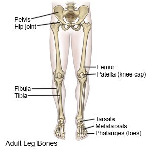
Tibia Shinbone Shaft Fractures Orthoinfo Aaos from orthoinfo.aaos.org It connects with the tibia and fibula bones of the lower leg. Also called the shin bone, the tibia is the longer of the two bones in the. The bones of the leg are the femur, tibia, fibula and patella.the foot bones shown in this diagram are the talus, navicular, cuneiform, cuboid, metatarsals and calcaneus. Some types of leg pain can be traced to problems in your lower spine. The patella is the kneecap and articulates with the distal femur. Now let's look at the tibia bone, which is the larger of the two leg bones, located medially. The knee joint is the largest joint in the body and is primarily a hinge joint, although some sliding and rotation occur. These are the femur, patella, tibia, fibula, tarsal bones, metatarsal bones, and phalanges (see figure 6.51).
The femur, or thighbone, is the longest and largest bone in the human body.
At the same time, the bones and joints of the leg and foot must be strong enough to support the body's weight while remaining. These muscles work together to produce movements such as standing, walking, running, and jumping. The lower limb contains 30 bones. This area is commonly referred to as the calf. Leg pain can also be caused by blood clots, varicose veins or poor circulation. The bones of the leg are the femur, tibia, fibula and patella.the foot bones shown in this diagram are the talus, navicular, cuneiform, cuboid, metatarsals and calcaneus. The hip itself is a ball and socket joint, much like the shoulder.the structures necessary to create this joint are the socket, the joint capsule, muscle, ligaments, and the neck. Bone anatomy of upper extremity 12 photos of the bone anatomy of upper extremity anatomy of upper limb bones pdf, bone anatomy of the upper limb, bone anatomy of upper extremity, bone anatomy of upper limb ppt, skeletal anatomy of the upper extremity, bone, anatomy. The muscles in the upper leg power many of our movements. The upper leg, in particular, is comprised of bones and muscles that are susceptible to injury, particularly when excess strain is placed upon them. The bones of the leg are the femur, tibia, fibula and patella.the foot bones shown in this diagram are the talus, navicular, cuneiform, cuboid, metatarsals and calcaneus. The femur is the single bone of the thigh. The talus is the bone at the top of the foot.
Share :
Post a Comment
for "Bones In Leg Diagram / Pin By Genna Hornsby On Anatomy Human Anatomy And Physiology Medical Anatomy Leg Anatomy"


Post a Comment for "Bones In Leg Diagram / Pin By Genna Hornsby On Anatomy Human Anatomy And Physiology Medical Anatomy Leg Anatomy"